ALERT!
This site is not optimized for Internet Explorer 8 (or older).
Please upgrade to a newer version of Internet Explorer or use an alternate browser such as Chrome or Firefox.
Five-on-a-Dice Port Technique for Robot-Assisted Right Upper Lobectomy for Hilar Lung Cancer
Kim, Min; Chan, Edward (2017): Five-on-a-Dice Port Technique for Robot-Assisted Right Upper Lobectomy for Hilar Lung Cancer.
CTSNet, Inc. https://doi.org/10.25373/ctsnet.5613001
Retrieved: 21:40, Nov 20, 2017 (GMT)
This video demonstrates the case of a 54-year-old woman with a 3.5 cm hilar mass abutting the main pulmonary artery, and located between the posterior ascending and superior segmental arteries.
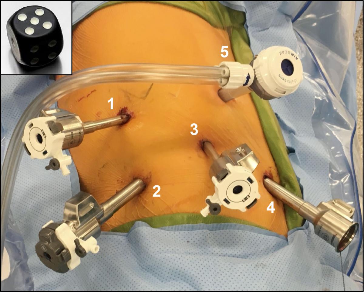
"Five-on-a-dice" refers to the appearance of the port placement of the four robot ports (labeled 1 - 4) and the assistant port (labeled 5). The authors find that this placement allows for easier dissection of hilar structures and the use of a robotic stapler during right upper lobe lobectomy.
Positron emission tomography/computed tomography showed localized disease with a standardized uptake value of 6.9. An endobronchial ultrasound showed no metastatic disease of the mediastinum, and magnetic resonance imaging of the patient’s brain was negative for metastatic disease. The patient underwent a robot-assisted right upper lobectomy and mediastinal lymph node dissection. A tip-up fenestrated grasper through port 1 was used to retract the lung. The Cadiere grasper and long bipolar grasper were used to dissect around the hilar structures through port 2 and port 4, respectively. The stapler was placed through ports 2 and 4 to divide hilar structures. A standard right upper lobectomy was performed successfully using this port placement and technique. The assistant was only used to remove lymph nodes and to place a tightly-rolled Ray-Tec sponge. The assistant was not needed for lung retraction or for firing a stapler around hilar structures. The patient was discharged home on postoperative day two. Her tumor’s final pathology showed a T2aN0M0/stage IB adenocarcinoma of the lung with negative margins.


