ALERT!
This site is not optimized for Internet Explorer 8 (or older).
Please upgrade to a newer version of Internet Explorer or use an alternate browser such as Chrome or Firefox.
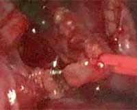
Ligation of the Thoracic Duct by a Posterior VATS Approach
This video demonstrates the ligation of the thoracic duct by a VATS approach. The patient suffered damage to the duct in the neck resulting in neck swelling and made local repair of the duct impossible. In this video the author demonstrates his technique for ligating the duct via a posterior VATS approach.
Left ventricular assist device (LVAD) implantation raises technical problems in patients with end stage heart failure and huge left ventricular aneurysm. This video shows how an LVAD (Jarvik 2000) can be implanted safely even in patients requiring left ventricular aneurysm resection.

The patient had a thyroidectomy and complete cervical lymph node dissection for thyroid carcinoma performed by the otolaryngologists 2 weeks previously. Unfortunately, the thoracic duct was damaged which resulted in severe neck swelling. Despite two attempts at re-exploration of the neck combined with a trial of a low fat diet, the neck swelling failed to diminish, and the effluent was clearly chyle. Of note there was no chylothorax.
He was referred for ligation of the thoracic duct. We elected to perform this by a posterior VATS approach, using the standard port arrangement that we use for posterior approach VATS lobectomy. This entails a first port anterior to latissimus dorsi in the 6th rib space. The second port is placed just posterior to the inferior tip of the scapula in the 5th intercostal space and this is used for the camera. In the mid axillary line in the 8th space a third utility port is used.
The lung was retracted anteriorly, and the pleura opened between the oesophagus and the azygous vein. An endo suture device was used to retract the sides of the pleura. The thoracic duct was then easily identified and triply clipped either side and cut. The specimen was removed and sent for histology which confirmed that this was the thoracic duct.
Paravertebral analgesia was then injected and the chest closed with a single 24 French drain that was removed later that day.


