Prior VATS lobectomy is a relative contraindication to ipsilateral repeat VATS lobectomy due to possible technical difficulties arising from adhesions and scarring, especially at the hilum. This video describes a VATS middle lobectomy in a 63 years old ex-smoker who in 2005 had a VATS right lower lobectomy for stage I squamous cell carcinoma. In 2008 he developed pulmonary tuberculosis of the right upper lobe that was successfully treated with anti-tuberculous medications. Then, in 2009 he underwent a successful right VATS middle lobectomy for a metachronous squamous cell carcinoma that was clinically stage I on CT and PET scan.
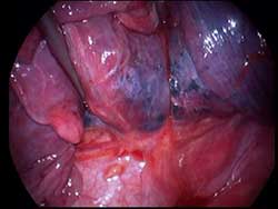
The original VATS incisions were reused. Chest wall and hilar adhesions were taken down meticulously. Lobectomy was accomplished by stapling the middle lobe vein first, followed by completing the fissures between upper and middle lobes. This helped in identifying the middle lobe pulmonary artery branches in the fissures. After ligating and doubly clipping the middle lobe pulmonary artery branches, they were divided. The right middle lobe bronchus was dissected and then divided with an endostapler. The bronchial stump was sealed with tissue glue. Operative time was 3 hours and 15 minutes. The hospital stay was 4 days and patient is well with no recurrence 12 months after surgery. The pathologic stage was T1N0M0, stage 1a.


