 |
 |
|
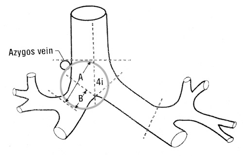
Fig.
52: Anatomic
landmarks for defining
N2 nodes in the lower
tracheobronchial tree.
The mediastinal nodes
that lie along the right
main bronchus and distal
to the cephalic border
of the azygos vein are
classified N2. The area
is depicted by "A" and
the length of the right
main bronchus is represented
by "B". These
distances are variable
with the length of the
main bronchus observed
between 0 cm., with one
or more segments of the
upper lobe arising from
the most distal trachea,
to 2.5 cm. The length
of the left main bronchus
is less variable and
the nodes along its length
are classified N2. |
|
|
Confirmation of N2 disease in patients
who meet all other criteria of operability
is recommended because all enlarged nodes
do not contain metastatic disease and the
positive predictive value of computed tomography
for identification of positive mediastinal
nodes is low15-16. In the absence of proof,
however, ipsilateral mediastinal lymph
nodes greater than 1 cm. in diameter are
staged N217. Similarly, pre- and retrotracheal
nodes that would be accessible to the surgeon
at thoracotomy are classified N2.
|