TX
If the primary tumor cannot be assessed,
a TX classification is assigned. The presence
of lung cancer cells in sputum with no
evidence of tumor by bronchoscopy or on
imaging (occult carcinoma) is classified
TX. A bronchioalveolar carcinoma that presents
as an infiltrate, with no evidence of tumor
or obstruction on imaging or at bronchoscopy
may be designated TX.
 |
 |
|
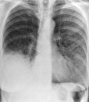
Fig.
1a: Posterior-anterior
chest radiograph showing
bronchioalveolar carcinoma
with nearly complete consolidation
of the right lower lobe
and extensive consolida
tion of the lingular segment,
TX. (Contralateral disease
would be designated M1) |
|
|
 |
|
|
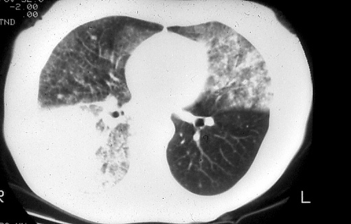
Fig.
1b: Computed tomographic
scan of the chest (lung
windows) shows the above
findings to greater advantage, TX. |
|
|
|