| « CTSNet Online Books | |
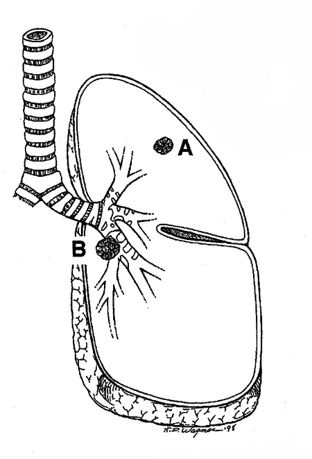
| LUNG
CANCER A Handbook for Staging, Imaging, and Lymph Node Classification by Clifton F. Mountain, MD; Herman I. Libshitz, MD; and Kay E. Hermes |
|
| Contents | About the Author(s) | Dedication and Acknowledgment |
|
||||||||||||||||||||||||||||||||
« previous
page | next page » |
| Copyright © 1999
- 2003 by CF Mountain and HI Libshitz, Houston, Texas. All rights
reserved. Printed in the United States of America by Charles P. Young Company. No part of this manual may be reproduced by any means without the prior written consent of the authors. |