T3
 |
 |
|
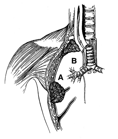
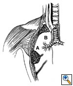
Fig.
10: T3: A tumor
of any size with direct
extension into the (a)
chest wall, including (b)
superior sulcus tumors,
or the diaphragm, mediastinal
pleura or pericardium,
without involving the heart,
great vessels, trachea,
esophagus or vertebral
body, or a tumor in the
main bronchus within 2
cm of the carina without
involving the carina. |
|
|
Limited, circumscribed, extrapulmonary extension
of the primary tumor is designated T3.
 |
|
|
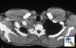
Fig.
11: Computed tomographic
scan of the chest showing
superior sulcus tumor in
the right apex with no
evidence of vertebral body
invasion, T3. |
|
|
|