ALERT!
This site is not optimized for Internet Explorer 8 (or older).
Please upgrade to a newer version of Internet Explorer or use an alternate browser such as Chrome or Firefox.
Anterior Chest Wall Resection and Identification of Externally Occult Lesions
Zhu AC, Bryan DS. Anterior Chest Wall Resection and Identification of Externally Occult Lesions. January 2024. doi:10.25373/ctsnet.25114352
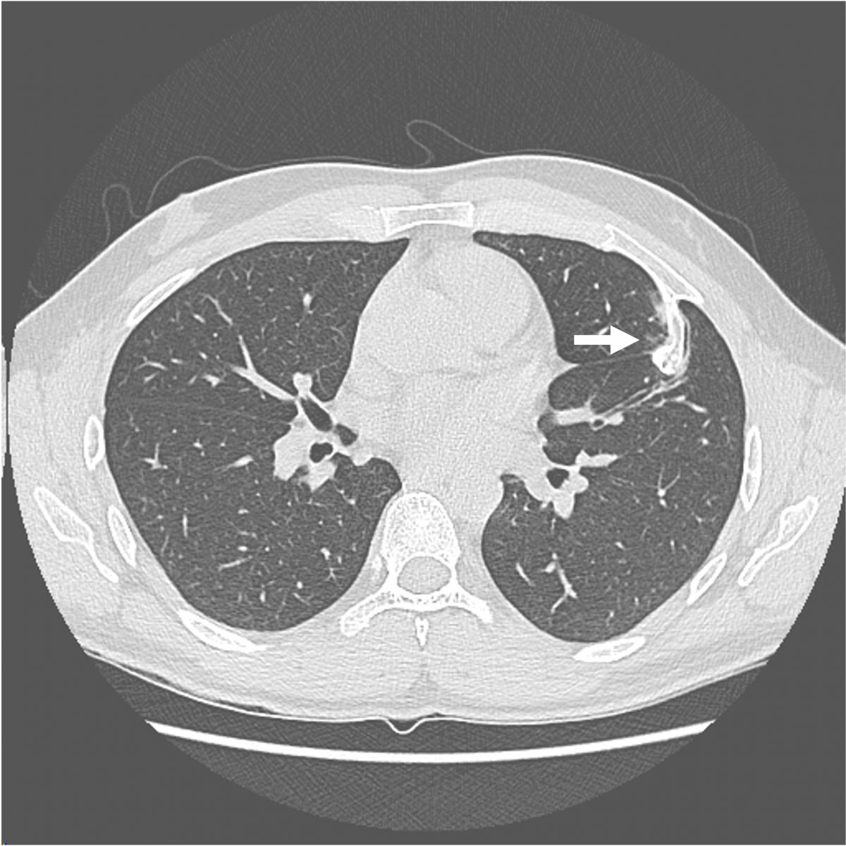
This video shows the case of a forty-year-old man who presented for evaluation of several months of anterior chest wall pain. Cross sectional imaging demonstrated a calcified anterior fourth rib lesion arising from the posterior table and projecting inward into the left hemithorax, measuring up to 3.8 cm (Figure 1). 
Physical examination was notable for an outwardly normal chest wall with no palpable abnormalities. The patient carried a history of multiple exostoses syndrome with multiple prior resections of long bone osteochondromas from infancy until 20 years of age (1). His family history was significant for multiple first-degree relatives with multiple exostoses syndrome.
While the lesion was without radiographic features commonly associated with malignancy, patients with multiple exostoses syndrome exhibit higher rates of malignant degeneration (2-4). Furthermore, the inward projection was thought to contribute to the patient’s chest wall pain. Thus, he underwent resection (5).
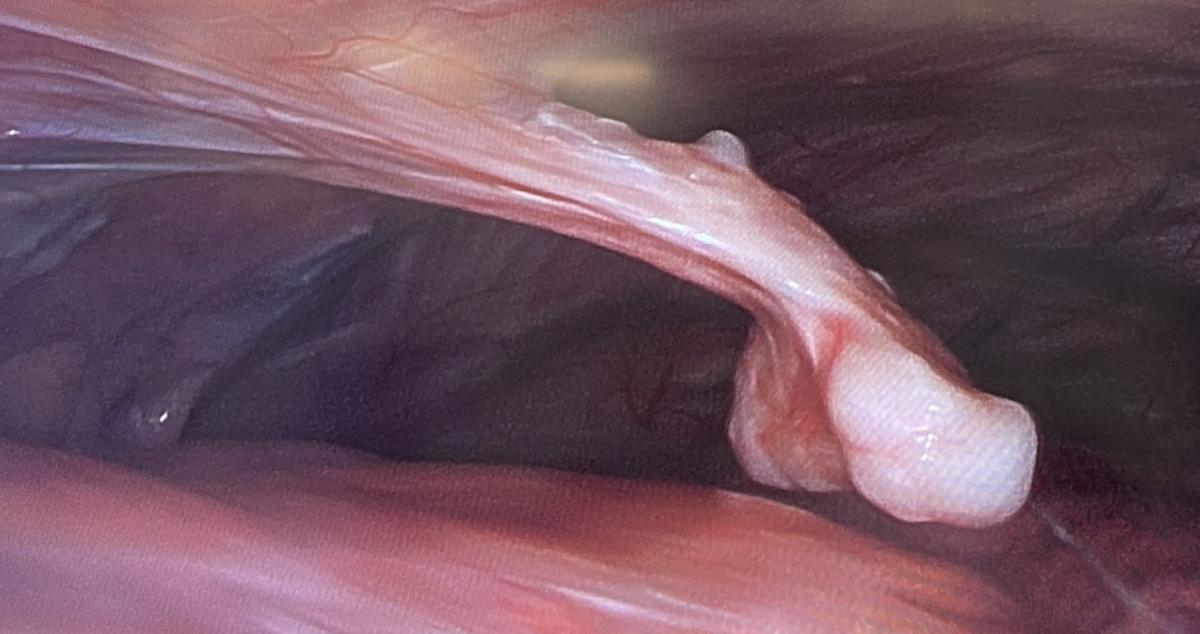
Surgeons elected to begin the operation by performing a thoracoscopic evaluation to localize the lesion and evaluate for any pleural adhesions. The patient was placed in a semi-right lateral decubitus position. An incision was made at the seventh intercostal space at the anterior axillary line and a thoracoscope was introduced. Inspection of the pleural space demonstrated a bony spur arising from the fourth rib (Figure 2). 
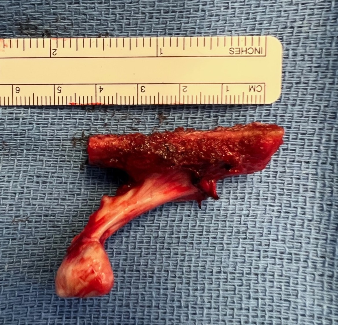
Surgeons made an inframammary incision, splitting the pectoralis fibers longitudinally and carrying the dissection down to the chest wall. A finder needle was used to identify the correct rib and the anterior and posterior transection points, minimizing the extent of the removal. Surgeons then performed a rib resection, electing to forgo chest wall reconstruction given the location and small size (Figure 3). The patient recovered well and was discharged home on postoperative day one.
References
- D'Arienzo A, Andreani L, Sacchetti F, Colangeli S, Capanna R. Hereditary Multiple Exostoses: Current Insights. Orthop Res Rev. 2019;11:199-211. Published 2019 Dec 13. doi:10.2147/ORR.S183979
- Levine BD, Motamedi K, Chow K, Gold RH, Seeger LL. CT of rib lesions. AJR Am J Roentgenol. 2009;193(1):5-13. doi:10.2214/AJR.08.1216
- Zuntini M, Pedrini E, Parra A, et al. Genetic models of osteochondroma onset and neoplastic progression: evidence for mechanisms alternative to EXT genes inactivation. Oncogene. 2010;29(26):3827-3834. doi:10.1038/onc.2010.135
- Rozeman LB, de Bruijn IH, Bacchini P, et al. Dedifferentiated peripheral chondrosarcomas: regulation of EXT-downstream molecules and differentiation-related genes. Mod Pathol. 2009;22(11):1489-1498. doi:10.1038/modpathol.2009.120
- Gonfiotti A, Salvicchi A, Voltolini L. Chest-Wall Tumors and Surgical Techniques: State-of-the-Art and Our Institutional Experience. J Clin Med. 2022;11(19):5516. Published 2022 Sep 20. doi:10.3390/jcm11195516
- Alabdullrahman LW, Byerly DW. Osteochondroma. In: StatPearls. Treasure Island (FL): StatPearls Publishing; February 5, 2023.
Disclaimer
The information and views presented on CTSNet.org represent the views of the authors and contributors of the material and not of CTSNet. Please review our full disclaimer page here.


