ALERT!
This site is not optimized for Internet Explorer 8 (or older).
Please upgrade to a newer version of Internet Explorer or use an alternate browser such as Chrome or Firefox.
Uniportal VATS Anatomic Right Basal Segmentectomy (S7-10)
Sesma J, Galvez C, Lin M-W, Nadal SB, Chen J-S. Uniportal VATS Anatomic Right Basal Segmentectomy (S7-10). January 2019. doi:10.25373/ctsnet.7591007.
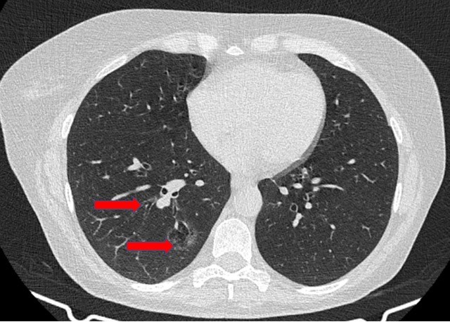
Anatomic sublobar resections have been used primarily for benign pulmonary lesions, lung metastases, and early stage lung cancer. There are many recent studies that support the oncological results of anatomic sublobar resections compared to lobectomy in terms of recurrence and overall survival [1-8]. In addition, the procedure’s performance through minimally invasive procedures is possible by experienced teams [9, 10]. The authors present a sublobar resection performed for a 50-year-old woman who was diagnosed with a 2.5 cm diffuse ground glass opacity adenocarcinoma in the right basal segments (Figure 1) that was identified during chronic asthma follow-up.
Preoperative studies ruled out nodal or distant involvement. A lung function test presented significant limitation, with an estimated postoperative DLCO near 30% after right lower lobectomy. Given the tumor characteristics, the absence of lymph node involvement, and the functional limitation, the authors decided to perform an anatomic segmentectomy of the basal segments (S7 + S8 + S9 + S10) through a uniportal video-assisted thoracoscopic (VATS) approach.
The authors began the procedure by making a 3.5 cm incision in the sixth intercostal space. A lymphadenectomy was performed in stations 12, 11, 10, 9, 8, 7, and 4R for intraoperative analysis. After confirmation of no nodal involvement, the major fissure was dissected, identifying the pulmonary artery and its branches to the basal segments and for segment 6 (S6). The common arterial branch for the basal segments was dissected and then divided using an endostapler. The inferior pulmonary vein was dissected, progressing distally until the vein of S6 was identified and the authors clearly found the division with the basal venous trunk. Then, the authors divided the basal venous trunk using an endostapler. The posterior fissure was completed with a stapler, identifying the venous branch for S6 from anterior and posterior in order to ensure its preservation.
The inferior lobar bronchus was dissected to the distal sublobar divisions, and the bronchus for the basal segments (B7-10) was dissected and divided with an endostapler after adequate ventilation of S6 was assured. Before this step, the authors also performed bronchoscopic evaluation of distal bronchial divisions. No intraoperative or postoperative complications were recorded. The chest tube was removed, and the patient was discharged home on postoperative day two. The final diagnosis was lepidic adenocarcinoma without nodal involvement (T2bN0M0).
Conclusion
The performance of anatomic segmentectomies through minimally invasive approaches as uniportal VATS is safe and feasible.
References
- Traibi A, Grigoroiu M, Boulitrop C, et al. Predictive factors for complications in anatomical pulmonary segmentectomies. Interact Cardiovasc Thorac Surg. 2013;17(5):838-844.
- Martin-Ucar AE, Nakas A, Pilling JE, West KJ, Waller DA. A case-matched study of anatomical segmentectomy versus lobectomy for stage I lung cancer in high-risk patients. Eur J Cardiothorac Surg. 2005;27(4):675-679.
- Date H, Andou A, Shimizu N. The value of limited resection for “clinical” stage I peripheral non-small cell lung cancer in poor-risk patients: comparison of limited resection and lobectomy by a computer-assisted matched study. Tumori. 1994;80(6):422-426.
- Okada M, Yoshikawa K, Hatta T, Tsubota N. Is segmentectomy with lymph node assessment an alternative to lobectomy for non-small cell lung cancer of 2 cm or smaller? Ann Thorac Surg. 2001;71(3):956-961.
- Watanabe T, Okada A, Imakiire T, Koike T, Hirono T. Intentional limited resection for small peripheral lung cancer based on intraoperative pathologic exploration. Jpn J Thorac Cardiovasc Surg. 2005;53(1):29-35.
- Campione A, Ligabue T, Luzzi L, et al. Comparison between segmentectomy and larger resection of stage IA non-small cell lung carcinoma. J Cardiovasc Surg (Torino). 2004;45(1):67-70.
- Keenan RJ, Landreneau RJ, Maley RH, et al. Segmental resection spares pulmonary function in patients with stage I lung cancer. Ann Thorac Surg. 2004;78(1):228-233.
- Dziedzic R, Zurek W, Marjanski T, et al. Stage I non-small-cell lung cancer: long-term results of lobectomy versus sublobar resection from the Polish National Lung Cancer Registry. Eur J Cardiothorac Surg. 2017;52(2):363-369.
- Galvez C, Lirio F, Sesma J, Baschwitz B, Bolufer S. Single-incision video-assisted thoracoscopic surgery left-lower lobe anterior segmentectomy (S8). J Vis Surg. 2017;3:114.
- Gonzalez Rivas, Lirio F, Sesma J. Uniportal anatomic combined unusual segmentectomies. J Vis Surg. 2017;3:91.


